Skin
The skin, covering most of the human body, is typically the primary focus for adornment in body modification. As the skin is the body's first defense against the remarkably hostile outside world, it is very tough and the body is extremely good at healing it. Furthermore, healthy skin has a tensile strength on the order of 15 lb/in², making and pulling possible.
Contents
Structure
On a basic anatomical level, skin is divided up into the epidermis (the skin that is constantly replacing itself and sloughing off) and the dermis (essentially permanent). Below that is the subcutaneous layer (fat cells) or hypodermis.
Epidermis
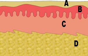
In the diagram above, layers A and B make up the epidermis, with A being the dead skins that line the surface, and B being living cells that move toward the surface, die, and fall off. Anything that ends up in this layer (such as tattoo ink, shallow piercings, and so on) will eventually be pushed out.
Dermis
Layer C is the dermis and is separated from the epidermis by a thin layer of connective tissue called a basement membrane. Stem cells at the top of the dermis seed cells into the epidermis above, but other than that this layer is static and does not exfoliate like the epidermis—thus when tattoo ink is placed in this layer with a tattoo machine, it stays for good. Piercings can migrate up or down given the right pressure, however, and contaminants can be carried away by the immune system. The dermis is structurally divided into two areas: a superficial area adjacent to the epidermis, called the papillary region, and a deep thicker area known as the reticular region. The papillary region is composed of loose (areolar) connective tissue. It is named for its fingerlike projections called papillae, that extend toward the epidermis. The papillae provide the dermis with a "bumpy" surface that interdigitates with the epidermis, strengthening the connection between the two layers of skin. In the palms, fingers, soles, and toes, the influence of the papillae projecting into the epidermis forms contours in the skin's surface. These are called friction ridges, because they help the hand or foot to grasp by increasing friction. Friction ridges occur in patterns that are genetically determined and are therefore unique to the individual, making it possible to use fingerprints or footprints as a means of identification. The reticular region lies deep in the papillary region and is usually much thicker. It is composed of dense irregular connective tissue, and receives its name from the dense concentration of collagenous, elastic, and reticular fibers that weave throughout it. These protein fibers give the dermis its properties of strength, extensibility, and elasticity. Located within the reticular region are also the roots of the hair, sebaceous glands, sweat glands, receptors, nails, and blood vessels.
Hypodermis
Below the dermis is the hypodermis (D) made up of loose connective tissue and fat cells. Deep wounds, such as those caused by a torn suspension hook, can expose this layer.
Notes
Like the rest of the world, skin contains massive amounts of bacteria, both inside and out. When a person washes their hands, they actually end up with more bacteria on the surface of the hands than before you washed. This is because by sloughing off the outer dead cells, normal flora naturally present in the skin are exposed. These organisms are largely harmless to the host, but not necessarily to others.
